The service was the first dedicated pleural service in the UK and is a tertiary referral unit.
We conduct a full range of pleural procedures, and run a dedicated Pleural Clinic several times per week, including in-clinic thoracic ultrasound.
We provide specialist assessment for patients with pleural disease, including:
- pneumothorax
- pleural infection
- cancer-related pleural effusion
- mesothelioma
- other pleural conditions.
We care for local patients and patients referred from other regions, providing a wide range of diagnostic tests and treatments for pleural disease. Our cases are discussed weekly at a multidisciplinary (MDT) meeting dedicated to pleural disease.
Pleural disease
Pleural disease occurs when there is a problem in the chest cavity, in the space between the lung and the chest wall.
This can result in the production of fluid in the chest cavity (called pleural effusion) which can lead to chest pain, breathlessness and other symptoms.
Fluid in the chest cavity may become infected (called empyema) and air may sometimes escape into the chest cavity (called pneumothorax).
There are over 50 recognised causes of pleural effusion, and our approach is to provide a service that accurately and quickly diagnoses the cause of the pleural problem so that treatments can be given.
Our team
Our service is run by:
- Dr Eihab Bedawi - Clinical Lead
- Professor Najib Rahman
- Dr Rob Hallifax
- Dr Ambika Talwar
- Dr Alastair Moore
- Dr John Park
- Dr Chia Ling Tey
along with two Clinical Fellows (Specialist Registrar level) in pleural disease, several dedicated Research Fellows, two Pleural Specialist Nurses and a Mesothelioma Specialist Nurse.
The team has extensive clinical experience and conduct research into the investigation, treatment and causes of pleural disease.
The service is supported by Radiologists with an expertise in pleural disease:
- Professor Fergus Gleeson
- Dr Rachel Benamore
- Dr Fiona MacLeod
- Dr Heiko Peschl
- Dr Victoria St Noble
- Dr Louise Wing
and, in addition, Pathologists and Oncologists who have a specialist portfolio.
There are excellent links between the Pleural Service and the Oxford Thoracic Surgery Service:
- Miss Elizabeth Belcher
- Mr Francesco Di Chiara
- Mr Dionisios Stavroulia
Our service
Patients referred from outside Oxfordshire are assessed and investigated in a minimum number of visits.
This includes a clinical assessment on the first visit which includes a thoracic ultrasound during the clinic, a second visit for specialist radiology (such as CT or PET scanning) and then an intervention / diagnostic procedure where needed.
Usually, clinical assessment in the dedicated Pleural Clinic will occur on Tuesday or Thursday, and interventions / diagnostic and treatment procedures are conducted on Mondays and Thursdays.
We aim to conduct the majority of investigations as day case or outpatient procedures, and then review one to two weeks later when the results are available.
We aim to see breathless patients for assessment within two weeks of referral.
Tests / procedures
We will nearly always need to do further tests to assess your condition and look into possible causes.
These include simple blood tests and X-rays. Other tests, which you may be less familiar with, include the following.
Thoracic ultrasound
This test uses ultrasound (as is used for scanning pregnant women) to carefully assess if fluid is present in the pleural cavity, and is the most accurate way of finding fluid in the chest.
We conduct this test during the initial clinic visit, and whenever an intervention is needed (see below) as it improves safety.
The test can be repeated during clinic visits to assess how much fluid is present, and does not involve any radiation.
Radiological tests
Beyond simple chest X-rays, the service uses Computed Tomography (CT) scans to assess most people with pleural disease.
This involves lying down in a large tunnel for a few minutes and creates a very accurate picture of what is happening inside the chest cavity. We may use a special sort of scan (PET) to assess if there are areas of chest cavity which are particularly 'active', which may lead to further tests.
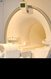
For patients with chest pain, we sometimes use MRI scanning of the chest.
Tests of pleural fluid
In patients with pleural effusion (fluid in the chest cavity), the most important test is to take a sample of the pleural fluid and test it in the laboratory.
This test is conducted in a highly sterile environment using ultrasound to guide where the sample should be taken. We always use local anaesthetic so that the test is not uncomfortable, and insert small tubes in to the chest cavity between the ribs. This allows us to take samples to be assessed in the laboratory.
If needed, we can drain a large amount of fluid which can relieve breathlessness and other symptoms.
More advanced tests
Sometimes the above tests do not establish the cause of the pleural condition, and in these cases a biopsy (small sample of the pleural lining) is required. This can be obtained in a number of ways, including using scans to guide a biopsy (ultrasound or CT), or using small cameras inserted in to the chest cavity using local anaesthetic and sedation to ensure comfort, called a thoracoscopy.
At thoracoscopy, we can take biopsies, fully drain the chest cavity and conduct a procedure to stop fluid coming back (called a pleurodesis) in a single procedure. Thoracoscopy establishes the diagnosis in more than 90 percent of cases.
In some cases, a full operation by our surgeons is required to obtain pleural biopsies.
Treatments
Treatment will depend on the cause of the pleural disease. However, draining of fluid or air and stopping it coming back are key parts of treatment, which help to relieve symptoms.
We can conduct this in a number of ways including using ultrasound to place small tubes in the chest, using the small camera in the chest (thoracoscopy) and even placing a tube in the chest which you can go home with and learn to drain yourself. This is very helpful as it allows us to treat some people as out patients if they do not want to stay in hospital.
The range of procedures offered by the unit include the following.
- Thoracic Ultrasound
- Diagnostic Pleural Aspirate
- Therapeutic Pleural Aspirate
- Intercostal drain insertion using guide-wire and blunt dissection
- Indwelling pleural catheter insertion, management and removal
- Pleurodesis using talc slurry and talc poudrage
- Diagnostic and Therapeutic Thoracoscopy
- Image-guided Pleural Biopsy (Ultrasound and CT-guided)
- Surgical Pleural Biopsy and Pleurectomy (via the Thoracic Surgery Service)
Research
The Oxford Pleural Unit is a research leader in the UK in the field of pleural disease and Professor Rahman is the Academic Lead.
We have conducted pivotal clinical trials in many areas of pleural disease, which have changed international guidelines and influence how patients are treated around the world, including:
- use of thoracic ultrasound
- optimal biopsy strategy
- safety of talc pleurodesis
- management of malignant pleural effusion
- use of indwelling pleural catheters
- novel treatments for pleural infection
- novel treatments for pneumothorax
- ambulatory care in pleural disease.
We design and deliver studies in pleural disease, and recruit to a number of studies including randomised and observational studies.
We may ask you to participate in research as part of your treatment, but this is always entirely your choice once we have explained it you, and your decision to participate will not affect the quality or timing of your care.
Contact us
If you would like more information about the Oxford Pleural Service, please contact:
Gill Radbourne
Email: gill.radbourne@ouh.nhs.uk
Resources
- Asthma + Lung UK
- British Thoracic Society
- Cystic Fibrosis Trust
- My Pleural Effusion Journey (information on management of cancerous pleural effusions)
- Mesothelioma UK
- Oxford Respiratory Trials Unit


