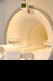
Xenon programme
ORRU is one of a few research groups in the UK actively taking part in imaging trials involving xenon.
We aim to improve both the quality of images for patients who have lung conditions and the treatment we are able to offer.
What is xenon?
Xenon is a colourless, odourless gas found in the Earth's atmosphere in trace amounts. It is currently used in hospitals as an anaesthetic. Within our research studies the xenon gas undergoes a process called 'hyperpolarisation' to make it more useful for imaging purposes. We have used xenon at the Churchill Hospital for many years in research studies.
What are the benefits of using xenon?
We are able to see the structure of the lungs and the air and blood flow and transfer of gas in great detail. We hope that xenon MRI scans will be available to patients in the future to allow us to detect differences in their disease, to determine how well they may respond to certain treatments and to be able to tailor treatments more effectively and appropriately.
Research studies
Updated June 2019
Xenon-Radiotherapy
We are recruiting participants who are receiving radiotherapy to the chest area. We want to look at the structure of the lungs, as well as the airflow and blood flow, in great detail. The purpose of this study is to see how xenon MRI pictures change with time before, during and after treatment.
Lung modelling using CT scans
ORRU is also developing a number of techniques that work alongside everyday scanning modalities (such as CT) to create 3D models of the lungs.
Artificial Intelligence in CT
More than 1.8 million cases of lung cancer are diagnosed each year worldwide. Early diagnosis of lung cancer is important to improve the outcome for the patient.
We currently use CT scan screening to detect small lung nodules before they become more aggressive. Recent studies have shown that looking at lung nodule characteristics (such as size, texture, growth rate, contrast enhancement) can also improve the accuracy of predicting the risk of cancer.
Optellum Ltd has developed a CT image analysis technique using machine deep learning models and large datasets that allows better nodule characterisation from a CT scan of the chest. The data from an earlier study has already shown that the software performs better than clinical experts.
The 'IDEAL Prospective' study aims to test the technique using CT scans from patients who are currently attending the CT department at the Churchill Hospital for investigation of a lung nodule. Patients are asked if they are happy for their images to be used, the images are then analysed by the algorithm which calculates a risk score to show how likely it suspects the lung nodule will turn out to be lung cancer.
This is one of the world's first prospective AI studies looking at current data rather than historical data.
As well as analysing the CT scan, we are also asking patients if they are happy to complete a number of questionnaires about their wellbeing and experience with the healthcare system.
One of our key objectives is to see whether a quicker diagnosis and treatment leads to an improvement in quality of life and a decrease in hospital visits and anxiety.
Contact us
If you would like to discuss these studies with us please contact us.
Email: radiology.research@ouh.nhs.uk
For more information please visit: