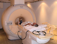
MRI scanner at the NOC
MRI is a very safe procedure. However, there are certain patients who cannot be scanned, for example those with a heart pacemaker.
Other implants might need to be checked before you are allowed in the scan room.
For this reason we ask all patients and visitors to fill in a safety questionnaire. This will normally be sent with your appointment letter.
If you answer yes to any of the questions, you must telephone the MRI Department and speak to a member of staff who will be able to give further advice. Please do this as soon as you receive your letter, as we may need to request information from other hospitals.
Radiology Department: 01865 738189
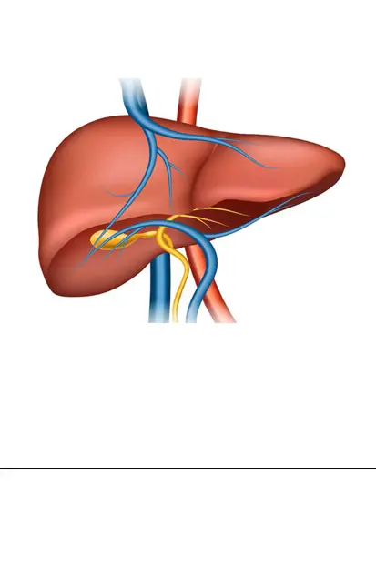
MRI provides detailed images of bone and soft tissues, such as lining of the joint, tendons and muscles. It shows up increased levels of inflammation and any tissue damage.
The scan takes approximately 20 minutes and requires the patient to lie still within the tunnel of the machine.
To avoid boredom, and block out some of the noise, young patients often listen to music via headphones or chat to their parents, who can sit at the end of the tunnel. Because of the need to lie very still some young children will have this scan under a general anaesthetic.
There are no X-rays involved. Instead images are taken using magnets, which means patients must not have metal objects with them. Sometimes a dye called gadolinium is injected via a cannula (tube) into the vein to help show up inflammation.
The MRI scanner uses harmless radio waves and a magnetic field to create images of tissues within the body. These images are more detailed than ultrasound and X-rays. MRI is painless and safe, although it is noisy and may create a feeling of claustrophobia.
In arthritis it is used to look at the lining of joints, the amount of fluid and whether there have been changes to the bony surface of the joint (called erosions). The MRI is also used to look at other tissues including muscles, ligaments and the brain and spinal cord.
How does MRI work?
The MRI scanner is like a big doughnut in shape, containing a large magnet within its ring and a long couch that passes through the hole. The patient lies on the couch and is carried into a tunnel. The noisy magnet then swings around the patient creating a magnetic field. This makes tiny particles within water molecules of the body (protons) point in one direction. Radio waves are then emitted to bounce off these protons and produce an image of the tissues being scanned.
Preparing for an MRI scan
Young children unable to keep still, or who will be frightened, will need a general anaesthetic. Your doctor will talk to you about this if you are parents or carers of a child who needs a scan.
Patients will be asked to wear a gown. The presence of a strong magnetic field means that metal objects are not permitted within the scanning room during a scan. These include:
- metal clips/zips on clothing and all jewellery
- medical equipment placed in the body, such as cardiac pacemakers, catheters with metal components, aneurysm clips and cochlear implants.
You will be asked about metal objects before a scan, so don't worry. In addition, females of childbearing age will be asked about the possibility of pregnancy because little is known about the effect of MRI scans on an unborn baby.
Going to the toilet is not possible during a scan, so patients are advised to limit the amount they drink from several hours before the scan, and to avoid coffee and tea.
Children can be accompanied by their parents/carers in the scanning room.
During an MRI
Once dressed in a medical gown, the patient is asked to lie down on a narrow table which then moves into the tunnel. An MRI scan lasts 30 minutes to an hour, depending on the type of scan. The patient must remain motionless, otherwise the picture is blurred and useless.
Throughout the procedure nurses, doctors and technicians are able to communicate with the patient via intercom.
Sometimes a dye may be injected via a drip into a vein. You will be told about this before the scan if this is going to happen.
Benefits of MRI
MRI is painless and safe. It provides detailed views of the body that may help with diagnosis or identify the extent of inflammation and/or any lasting damage. This helps to determine treatment.
Risks of MRI
Please inform the technician or doctor if there is a risk of pregnancy or metal objects within the body. If a dye is given there is a small risk of allergic reaction.
After an MRI scan
The radiologist, a doctor experienced in MRI and X-rays, will analyse the images and write a report interpreting the scan. The MRI results are then passed back to the referring doctors, who will explain them to you.
Specialised types of MRI
- Magnetic Resonance Angiography (MRI) - to look at blood vessels.
A contrast dye is injected, and helps the blood vessels to show up brighter. - Functional MRI - to look at joints and the brain.