Departments and services
You can view our departments and services alphabetically. Some areas also have dedicated microsites, which are featured in the linked images below.
You can view our departments and services alphabetically. Some areas also have dedicated microsites, which are featured in the linked images below.
Telephone number 0300 304 7777
John Radcliffe Hospital
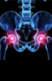
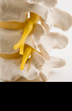
Nuffield Orthopaedic Centre
Churchill Hospital
Horton General Hospital