Can MRI scans be used to assess kidneys before and after transplantation?
23 January 2026
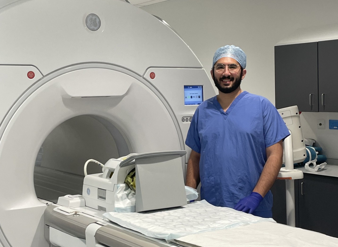
Dr Mohamed Elzawahry on the occasion of the first scan
A new clinical study is exploring whether advanced MRI scanning of kidneys before and after transplantation could provide a non-invasive, objective way to evaluate donor organs and monitor transplant health.
The study, led by researchers at the University of Oxford, Oxford University Hospitals (OUH) and the University of Nottingham, could potentially transform how kidneys are assessed and managed in routine clinical practice.
Due to the limited pool of kidney donors, transplant teams are increasingly accepting kidneys donated by older donors and donors with multiple medical conditions. This can affect the function and longevity of the transplanted kidney. Predicting how well donor kidneys will function after transplantation is therefore crucial, yet accurate assessment tools are currently lacking.
Could MRI scans of kidneys before and after transplantation provide an objective and accurate assessment tool that can be embedded into the transplant pathway and ultimately become part of routine clinical practice?
This is what Professors Maria Kaisar of the University of Oxford’s Nuffield Department of Surgical Sciences, and Susan Francis, of the University of Nottingham, aim to find out.
The ADMIRE project team, funded by the Stoneygate Trust and Garfield Weston Foundation, and supported by Kidney Research UK, has launched this first-in-human clinical study of ex vivo MRI scanning of donor kidneys prior to transplantation, followed by a three-month post-transplant MRI scan of recipients. The study is being conducted at OUH’s Oxford Transplant Centre.
Dr Mohamed Elzawahry, Clinical Study Associate Principal Investigator, Nuffield Department of Surgical Sciences, has recruited the first donor kidney that was scanned and subsequently transplanted, under the supervision of Dr Edward Sharples Principal Investigator at OUH.
The aim of the study is to determine whether this non-invasive assessment can be feasibly incorporated into clinical practice to evaluate donor kidneys and monitor transplant recipients.
The ADMIRE project is also investigating early, non-invasive monitoring of kidney transplant recipients, without the need for biopsies. This approach could enable earlier detection of transplant dysfunction and create a window of opportunity for intervention, helping transplanted kidneys to function for longer.
David Crosby, Chief Research Officer at Kidney Research UK, commented: “There are around 7,000 people currently waiting for a kidney transplant in the UK. We need to find new ways of increasing the number of kidneys available for transplant and to make them function better for longer.
“This new approach using MRI scans could allow doctors to better predict how well a donated kidney could function in the recipient and to continue to monitor this after transplant. This could mean healthier kidneys can be transplanted which last longer. More research is needed to develop this concept, and we look forward to the next steps.”
In parallel, the ADMIRE team, in collaboration with Professor David Long of University College London, is studying deceased donor biopsies and three-dimensional histological patterns to improve the interpretation of MRI findings in clinical practice.
Successful completion of this study could lead to a future clinical trial in which MRI-based assessment of donor kidneys and transplant recipients informs clinical decision-making and potentially improves transplant outcomes.